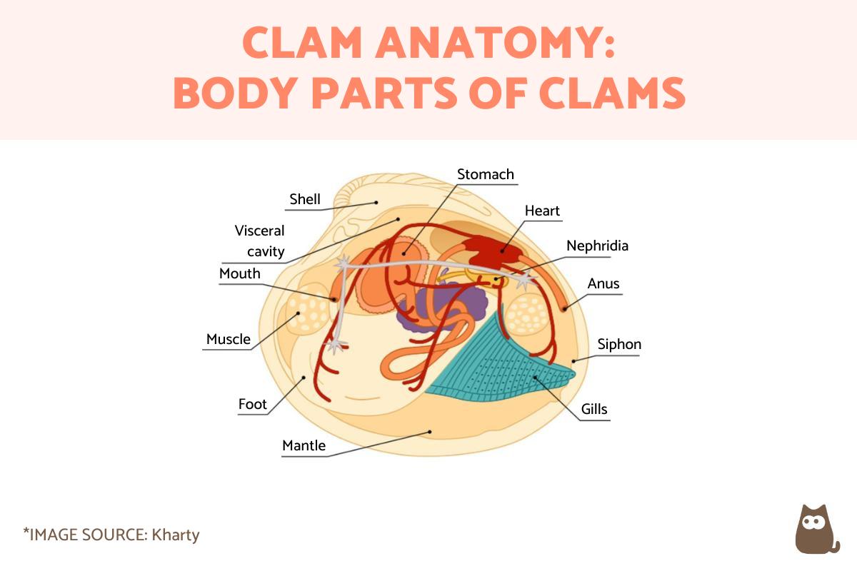
Clam Anatomy - Parts of a Clam


Clams are bivalve mollusks that play an essential role in aquatic ecosystems. They contribute to their habitats not only by providing a food source for other animals, but also in water filtration, nutrient recycling and other important benefits. They can do so thanks to a specialized anatomy which is typical of other bivalves. In addition to their characteristic protective shell, they also have internal organs made of soft tissue which allow the clam to survive in their habitat.
AnimalWised explains more by looking in depth at clam anatomy. We provide a list of the body parts of a clam with a diagram and photos to help you recognize them better.
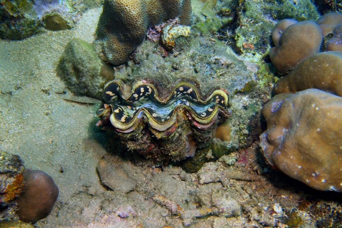
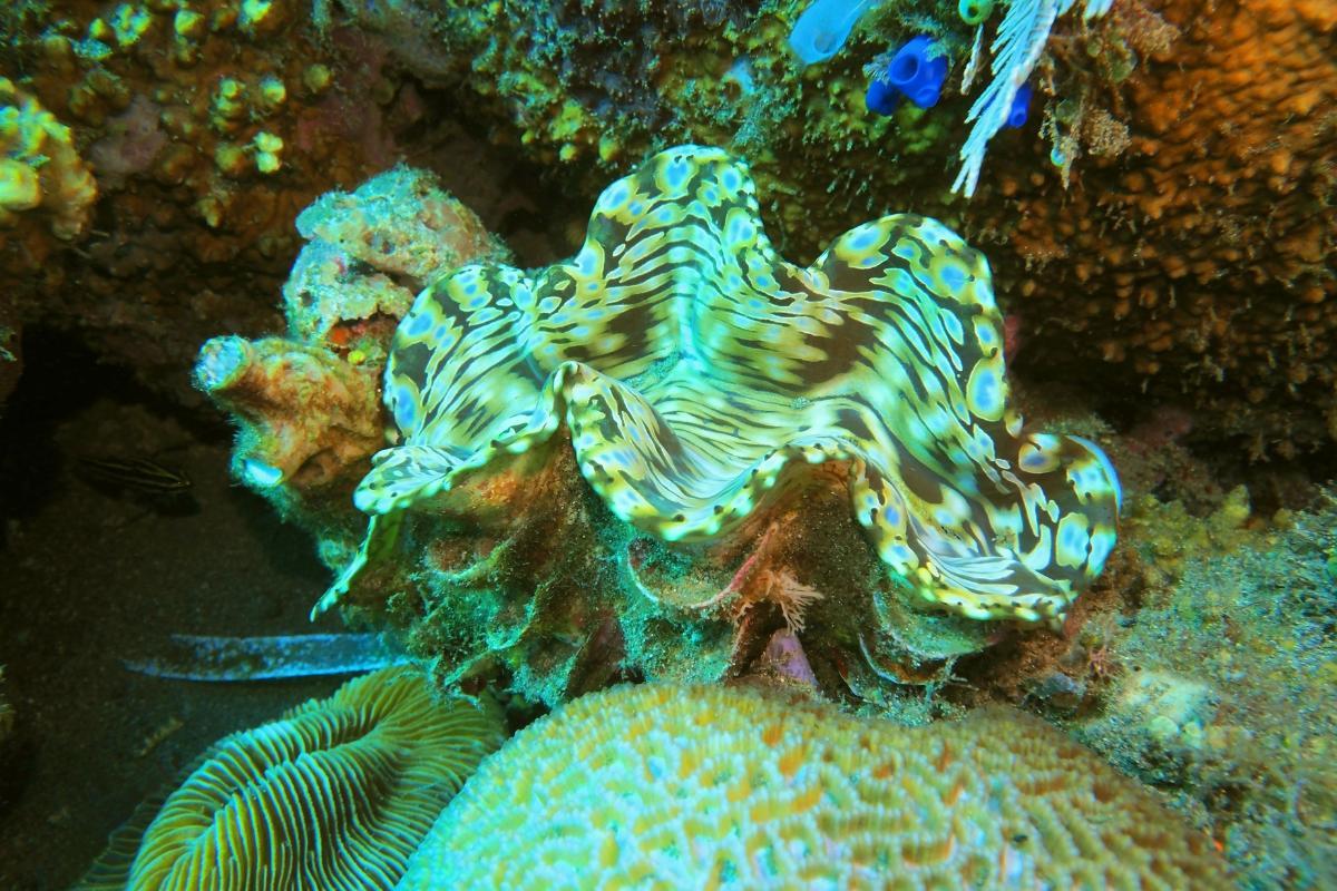
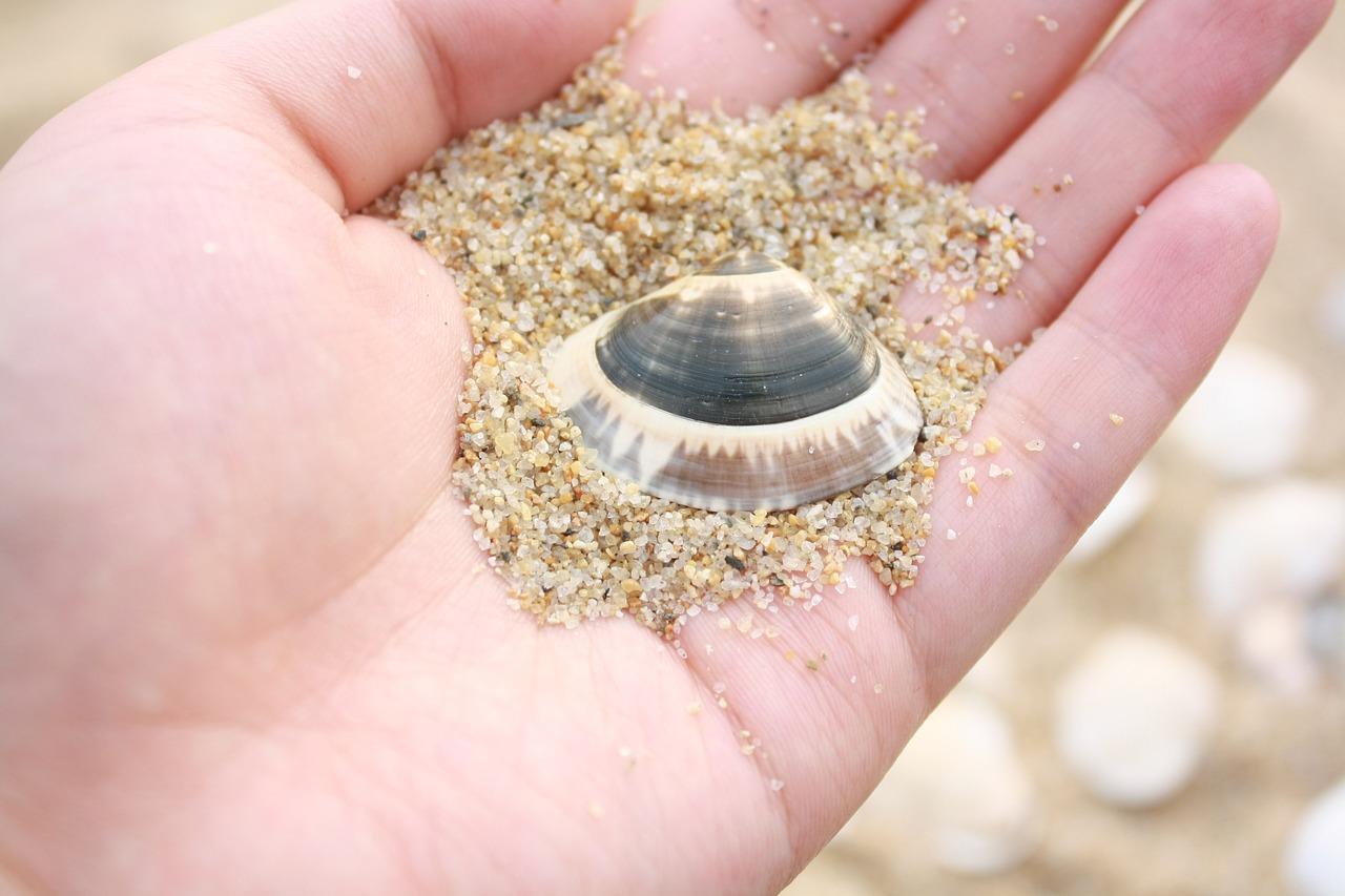
Shell (valve)
The shell of the clam is made up of two valves, each of which is joined together by a hinge. These valves are composed of concentric layers of calcium carbonate, providing a robust and protective structure. Since it is known for its valves, the shell is scientifically known as a valve. The shells can vary in shape, size and color, depending on the species of clam. The parts of a clam shell are as follows:
- Periostracum: it is the outermost layer of the shell, composed mainly of proteins. Its function is to protect the inner layers of the shell from physical damage and biological attacks, as well as prevent erosion.
- Prismatic layer: this layer is located below the periostracum and is made up of calcium carbonate crystals arranged in the shape of prisms. It provides the main structural strength to the shell.
- Nacre (mother of pearl): the nacre is the innermost layer of the shell and is composed of thin layers of calcium carbonate arranged in an orderly manner. This layer is smooth and shiny and may have an iridescent finish. The nacre is also the layer used in the formation of pearls in some clam species.
The shell serves as a defensive barrier against predatory animals, adverse environmental conditions and physical damage from any kind of trauma. It allows the clam to protect its soft inner tissues which are much more vulnerable. Additionally, clam shells help to maintain moisture in these soft tissues, something especially important when the clam is exposed to dry conditions or when in low-water environments.
Discover the anatomy of another famous animal with a shell in our article explaining the parts of a turtle.
Mantle
The mantle is a thin membrane that covers the body of the clam and is attached to the inner edge of the shell valves. It extends along the edge of the valves and forms a sac containing the soft body of the mollusk. This membrane is composed of a layer of specialized epithelial cells that have several important functions.
Some of the characteristics and functions of this part of the clam are:
- Secreting material which creates the shell: these epithelial cells are responsible for the deposition of the layers of calcium carbonate that make up the shell of the clam. The space between the mantle and the shell valves is known as the mantle cavity. It fills with water that the clam uses for various physiological functions.
- Formation and maintenance of the shell: by secreting calcium carbonate and proteins, the mantle allows the construction and growth of the shells. This ensures the shell continues to protect the soft body as the clam grows.
- Facilitates the circulation of water: it does so through the mantle cavity using siphons. This circulation is essential for gas exchange and food filtration. Water is drawn in through the inhalation siphon, passes through the gills for gas exchange and filtration and is then expelled through the exhalation siphon.
- Protection: it acts as a protective barrier for the soft body of the clam, covering and protecting delicate internal organs. It also helps keep the soft body free of sediment particles and microorganisms that could cause infections.
In case of damage or injury to the shell, the mantle can repair and regenerate the affected areas. This healing process is crucial to maintaining the structural integrity of the shell and protecting the mollusk.
Learn more about another animal that is capable of regeneration with our article asking is the starfish a vertebrate or an invertebrate?

Hinge
The hinge is made up of adductor muscles, which are responsible for the movement of the valves. These muscles are located in the internal region of the concha and are divided into two pairs:
- Anterior adductor muscles: located at the front of the clam, these muscles help close the valves.
- Posterior adductor muscles: located at the back of the clam, they also help close the valves and provide additional stability.
These muscles are connected to the shell by ligaments and other connective tissues. The hinge performs functions such as controlling the opening and closing of the valves. It does so through the contraction and relaxation of the adductor muscles. When the adductor muscles contract, the valves close. When they relax, the valves open. This mechanism allows the clam to regulate the amount of shell opening.
The hinge also acts by allowing the clam to close its valves tightly as a defense against predatory animals. This quick and firm closing ability is crucial to protecting the clam's internal soft tissues.
The opening and closing of the valves also helps the clam adapt to its environment. By opening the shells, the clam can filter water to feed and breathe. By closing them, they can protect themselves from adverse environmental conditions, such as low oxygen concentrations or desiccation. In clams that can swim, the hinge plays a part by rapidly clapping the shell together to propel it through the water.
Siphon
The siphon in clams is a tubular structure that plays a crucial role in the circulation of water within the mantle cavity. It is made up of two main parts:
- Inhalant siphon: a thin tube that extends from the mantle cavity outward. It is designed to introduce water from the environment into the interior of the clam. It allows water rich in nutrients and oxygen to enter the clam's mantle cavity. As water flows through the mantle cavity, the gills filter food particles and microorganisms present in the water. Oxygen dissolved in the water is absorbed by the gills for respiration, while carbon dioxide produced by the clam is released into the water as waste product.
- Exhalant siphon: similar to the inhalant siphon, it is a tube that expels water once it has passed through the mantle cavity. It expels water after it has passed through the gills and has been used for filtration and respiration. In addition to water, the exhaling siphon removes metabolic waste and particles that were not retained by the gills.
The siphons can extend and retract, allowing the clam to adjust its position in the substrate or respond to changes in the environment. Such changes include the arrival of predators or changes in water quality. When the clam feels threatened, it can retract the siphons and close the valves tightly to protect itself.

Gills
We may associated gills with larger animals such as various types of fish, but clams have gills in much the same way. They are the structures crucial for underwater respiration, as well as for other processes such as feeding. They are located in the mantle cavity and are essential for survival in all types of bivalve mollusks.
Clam gills are formed by thin, lamellar filaments in the mantle cavity. They have specialized cells for gas exchange and food capture. Their structure optimizes the capture of oxygen and the elimination of carbon dioxide.
The gills are one of the parts of clams that have two different functions:
- Gas exchange: they extract dissolved oxygen from the water flowing through the mantle cavity. Oxygen is absorbed by the gill tissue and transported to the clam's circulatory system. Carbon dioxide is a metabolic waste product that diffuses from the clam's blood into the water in the gills to be expelled through the exhalant siphon.
- Filtration: their filter by capturing small food particles suspended in the water, such as phytoplankton and zooplankton. Particles trapped in the gills are transported by a mucus that coats the gill filaments to the clam's mouth for digestion.
The gills help regulate the flow of water through the mantle cavity. They ensure that water flows efficiently to maximize nutrient capture and gas exchange.
Foot
The foot in clams is a fundamental muscular structure that plays several important roles. It is a soft, extended muscle mass found at the base of the clam's soft body, the reason it is known as a foot. It extends from the underside of the mantle and is composed of highly flexible muscle tissue.
The clam foot allows it to burrow into the bottom sediment of its aquatic habitat. By contracting and expanding its foot muscles, the clam can move sediment around it to bury itself. This ability to burrow provides a form of shelter and protection.
Once the clam has buried itself, the foot helps anchor it firmly in position. The clam can expand its foot to better secure its hold in the substrate, preventing it from being carried away by water currents or other disturbances in the seabed.
Although clams are mostly sessile animals (i.e. they do not actively move), the foot allows them to make limited movements on the substrate. By contracting and expanding the foot, clams can move slowly in their environment to search for food or change positions.
The foot can respond to environmental conditions, such as changes in the substrate or the presence of predators. If the clam detects a threat, it can use its foot to bury itself deeper or change its position to improve its safety. Additionally, as it burrows into the sediment, the clam foot can disturb the substrate around it, helping the clam access areas of water with higher concentrations of nutrients.
Internal organs
The internal organs of clams are located in the visceral cavity. This is a space within the mantle cavity that houses the systems essential for the life of the mollusk.
Heart
The heart of the clam is a small and relatively simple organ, located in the center of the visceral cavity . It pumps hemolymph (similar to blood) through the clam's open circulatory system. Hemolymph transports oxygen and nutrients to tissues and removes metabolic wastes. The heart also regulates internal pressure and hemolymph flow.
Discover more about hemolymph in invertebrates with our article explaining whether insects have blood.
Digestive system
The parts of the clam digestive system include:
- Mouth: located in the anterior part of the body and is the entry point for food filtered from the water.
- Esophagus: a tube that connects the mouth to the stomach, transporting food to the digestive system.
- Stomach: where the digestion of food begins. In some clams, the stomach is divided into several chambers for more efficient digestion.
- Intestine: a long tube that extends from the stomach to the anus. In the intestine, nutrients are absorbed and waste is prepared for elimination. The anus is the exit point for indigestible waste from the digestive system.
Reproductive organs
Known as gonads, the reproductive organs are those that produce gametes (eggs or sperm). In clams, the reproductive organs are located near the stomach and intestine. Eggs or sperm are released into the water through the mantle cavity during the reproduction process. Fertilization can occur in the water or inside the clam, depending on the species.
Nephridia
Nephridia are excretory organs located in the visceral cavity. They can be considered the equivalent of vertebrate kidneys in invertebrate animals. They filter metabolic waste from the hemolymph and eliminate it through the mantle cavity. They help maintain the balance of salts and the concentration of internal fluids.
Circulatory system
Clam anatomy has an open circulatory system, meaning the hemolymph flows directly over the internal organs instead of being contained in blood vessels. It transports nutrients, oxygen and waste through the hemolymph, helping in the distribution of substances essential for the functioning of the body and in the elimination of waste products.
If you want to learn more about the body parts of other aquatic invertebrates, you may want to check out our article on the anatomy of a crab.

Labial palps
Labial palps are sensory structures located near the mouth of clams. They are soft, elongated appendages that extend from the edge of the oral cavity. They have an elongated, flexible shape and their number and arrangement can vary between different species of clams.
The labial palps help collect and direct food particles that have been filtered by the gills. These appendages touch and manipulate food to ensure it reaches the mouth. Food particles are directed towards the mouth with the help of the movements of the labial palps, which act as guides and transporters.
Basically, labial palps have tactile and chemical sensors that allow the clam to detect the presence and quality of food particles. This helps ensure that only appropriate foods are eaten. They help separate desirable foods from waste and inedible particles, contributing to efficient water filtration and nutrient processing. Essentially, they allow the survival of the clam.
Discover more about the anatomy of marine animals with our article on shark body parts.
If you want to read similar articles to Clam Anatomy - Parts of a Clam, we recommend you visit our Facts about the animal kingdom category.
- da Costa, F. (2012). Introduction to the biology of clams. Clam Fisheries and Aquaculture, 1.
- Hanks, R. W. (1963). The soft-shell clam (No. 162). The Bureau.
- Korniushin, A. V. (1995). Anatomy of some pill clams (Bivalvia: Pisidioidea) from Africa, with the description of new taxa. Journal of molluscan studies, 61(2), 163-172.
- Smolowitz, R. (2021). Mollusca: Bivalvia. Invertebrate Histology, 163-183.


